Staining and Microbiologic Features:
- C. Jejuni is an S-shaped rod that is non-spore forming and exhibits gram-negative staining [1].
- C. Jejuni flourishes in an environment with lowered oxygen levels (5-7%) and heightened carbon dioxide levels (5-10%), with optimal growth observed at 42°C compared to 37°C [2].
- Unlike other gram-negative bacteria that exhibit cell wall lipopolysaccharides (LPS) with endotoxin activity, C. jejuni expresses lipooligosaccharides.
- It tests positive for oxidase and catalase [3].
- The single polar flagellum of C. jejuni helps in its motility [1].
- Reservoirs for C. Jejuni include poultry and wild domestic animals [4].
- C. Jejuni is considered acid-sensitive, making it susceptible to the acidic conditions of the stomach. Consequently, it requires a high infective dose (about 104organisms) to initiate an infection in the intestine [3].
Virulence:
- Similar to E. coli, C. Jejuni produces enterotoxin (LT toxin) [5].
- It produces cytotoxin that damages the intestinal mucosa [5].
- H antigen is another important virulence factor for C. Jejuni [4].
Transmission:
- Transmission occurs through the fecal-oral route [4]
- Consumption of unpasteurized milk and uncooked poultry [4]
Diseases and Complications:
- Diarrhea: The patient can present with fever, headache, abdominal cramps, and bloody diarrhea [6].
- Some strains of C. Jejuni (especially serotype O:19) are associated with Guillain-Barré Syndrome. Antibodies produced during infection cross-react with peripheral nerves and lead to autoimmune-induced demyelination, resulting in Guillain-Barré Syndrome (GBS) symptoms [7].
- Some patients (particularly with HLA-B27 phenotype) can also develop Reactive Arthritis after C. Jejuni infection [7].
Diagnostic Testing:
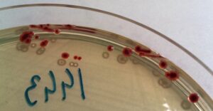
Campylobacter jejuni colonies” by Stefan Walkowski is licensed under CC BY-SA 4.0.
References:
1 Jawetz, Melnick, & Adelberg’s Medical Microbiology Twenty-Seventh Edition (page no: 256)
2 Medical Microbiology by Patrick R. Murray Ph.D., Ken Rosenthal Ph.D., Michael A. Pfaller MD, 8th edition (page no: 281)
3 Jawetz, Melnick, & Adelberg’s Medical Microbiology Twenty-Seventh Edition (page no: 257)
4 CMMRS edition 6, 2016-17 (page no: 86)
5 CMMRS edition 6, 2016-17 (page no: 81)
6 CMMRS edition 6, 2016-17 (page no: 80)
7 Medical Microbiology by Patrick R. Murray Ph.D., Ken Rosenthal Ph.D., Michael A. Pfaller MD, 8th edition (page no: 282)
8 Medical Microbiology by Patrick R. Murray Ph.D., Ken Rosenthal Ph.D., Michael A. Pfaller MD, 8th edition (page no: 283)
#Microbiology #Campylobacter #Jejuni


More Stories
COVID Therapeutics
MSRA: Complete Guide 2024
How to lose your compassion: the brutal reality of medical internship